Nowadays, 3D technologies have been increasingly used in many fields of medicine. Tests have already been done to produce bone cartilage for the kneecap, nose and ears. Also, scientists are working to make 3D-bioprinted artificial eyeballs. This is difficult, but if successful, it will become possible to restore vision to blind people. All evidence suggests, that soon 3D bioprinters will be capable of printing whole cells from human organ stem cells.

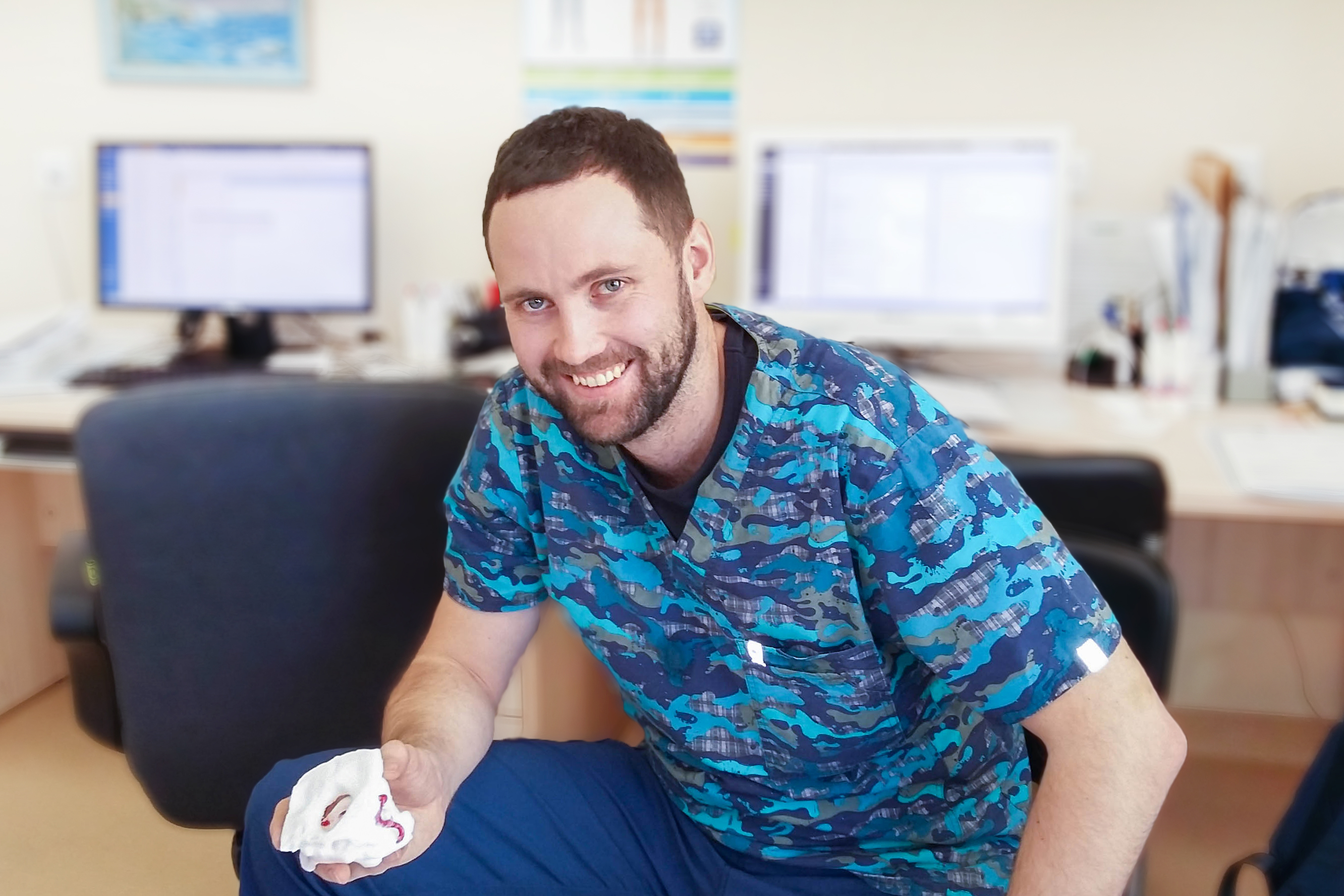
Roman Kovalenko, neurosurgeon
But there’s more. 3D technologies can be used in many more ways. They can help doctors create excellent tools to perform complex medical procedures and even surgery.
Recently, Roman Kovalenko, a neurosurgeon from Almazov Centre, won the best research paper contest held among young neurosurgeons, neurologists, neuroradiologists and other neurology physicians with an article “Using personalized 3D-printed navigation templates for transpedicular screw fixation in the cervical spine: the results of a prospective two-center pilot study”.
Today, often with spinal disorders, neurosurgeons have to resort to surgery when special screw systems are inserted into the vertebrae of a patient. Since vascular and neural structures are located very close to the vertebrae, implants should be inserted with high precision and on a preset path. In some cases, it is quite difficult. For example, in scoliotic deformities, congenital anomalies and neoplastic lesions the anatomical structure of the vertebrae can be changed. It leads to the risks of complications in the insertion of screws (e.g. damage to arterial structures).
One of the classic techniques used by surgeons to improve the accuracy and safety of implantation is an intraoperative x-ray. However sometimes an x-ray projection is not enough for surgeons, they need to see a three-dimensional image in order to properly implant the screw. Until recently, a more accurate navigational method was the use of intraoperative CT and navigation system. CT is performed on the patient during the surgery. The images are then linked to a navigation system, and the surgeon uses this special navigation. This increases the quality of implantation and reduces the rate of complications. This equipment is quite expensive, and the patient is exposed to a large radiation load during surgery.
In his article, Roman Kovalenko described an alternative method for the precise introduction of fixation screws into the spine.

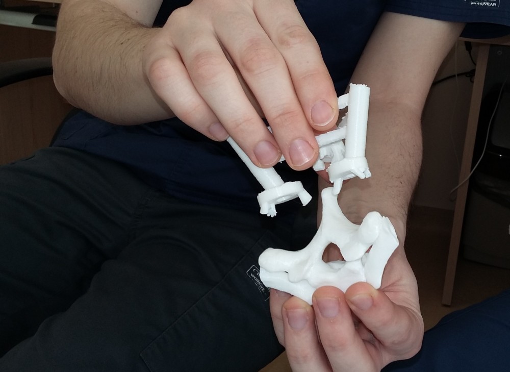
Vertebra model and 3D-printed navigation template
The method is based on the use of 3D-printed templates fitting with the patient's vertebra to ensure the perfect insertion of screws. The rapid prototyping technique (3D printing) has recently been broadly introduced in various areas of surgery.
“The method enables precise fixation of screws in cervical spine surgeries, thus reducing the rate of complications and the burden of repeated surgery. Other advantages are the significant reduction of radiation load on the patient (since there is no need for images in different projections), ease of use and relatively low cost. This is a very efficient solution,” the neurosurgeon noted.
On the initiative of Roman Kovalenko, this technique is now used in two Russian centres: Almazov Centre and Vreden Russian Research Institute of Traumatology and Orthopedics. The neurosurgeons of Almazov Centre began to use this method in October 2017. Over this time, the surgery has been performed in 26 patients.
