Doctors at Department of Pediatric Neurosurgery No. 7 of Almazov Centre successfully use additive technologies. The 3D printing technology has been in use since 2017 for surgical procedures in children with complex concomitant anomalies of the skull, tumors of the skull bones and complex trephine defects. Preoperative preparation makes it possible to model implants before the procedure, based on the individual’s anatomy, and for skull base and orbital tumors – to plan beforehand the course of the surgical access and the minimum craniotomy area. This greatly reduces the invasiveness of surgery and improves functional and cosmetic outcomes.
One of the cases involving the use of this technique was a teenager who underwent surgery at the end of 2020. The patient presented with proptosis (bulging eyes) and pain in the left superciliary area.
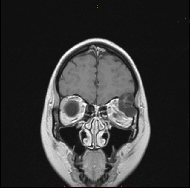
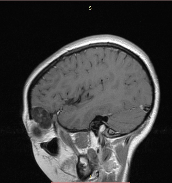
Examination and imaging revealed a left orbital mass spreading to the orbit walls and cranial cavity.
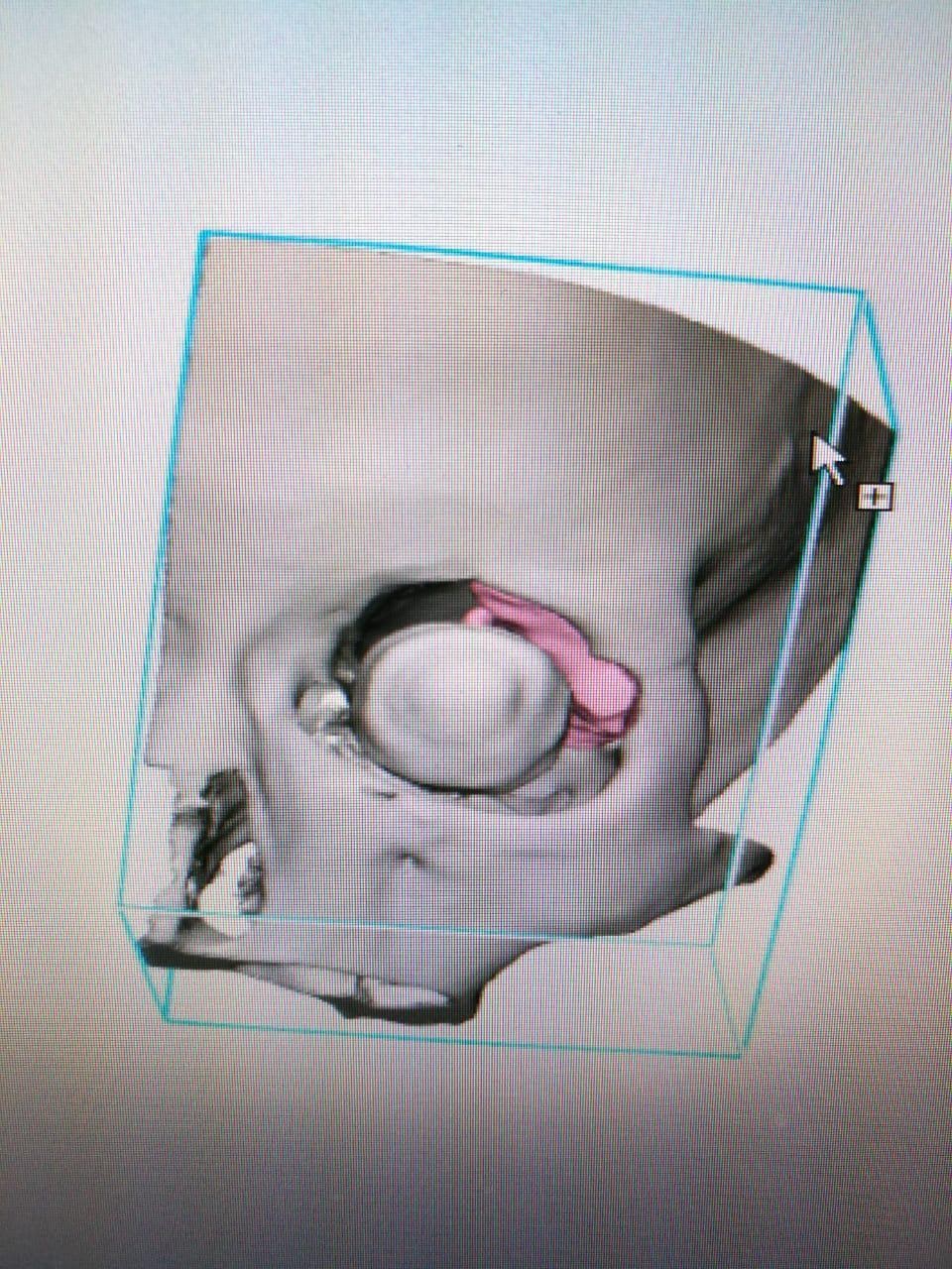
“We used modern neuroimaging methods followed by the construction of a virtual model to create a real-size 3D model of the tumor and surrounding structures. Such a thorough preoperative preparation with planning for determining the course of surgical actions, bone dissection lines and the minimum extent of resection of the affected structures made it possible to perform minimal access surgery,” said pediatric neurosurgeon Vadim Ivanov.

3D reconstruction of skull bones before surgery. Tumor destroying the orbital roof and invading the zygomatic process of frontal bone
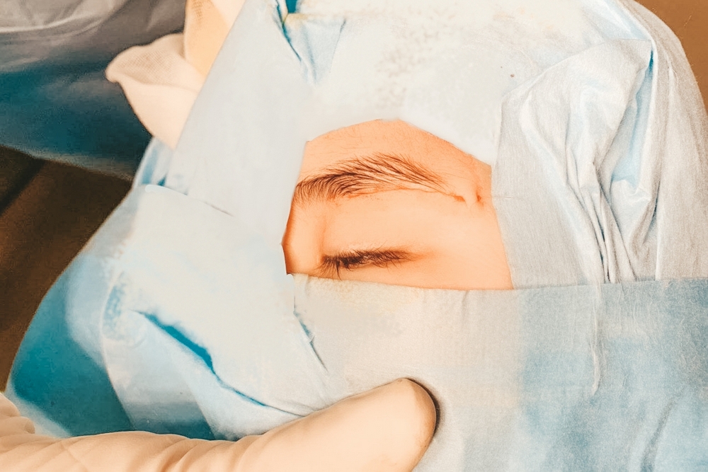
The doctors performed a minimally invasive surgery to remove the skull base tumor using additive technologies (3D printing). The surgery lasted 90 minutes. During this time, the specialists performed osteoplastic resection of the zygomatic process of the frontal bone, removed the mass and the affected structures. The bone flap was then reinserted and fixed with biodegradable miniplates. The cosmetic suture will make the subsequent scar almost invisible.
 |
 |
 |
| MRI before surgery | 3D modeling before surgery The mass is marked in pink |
|
Since 2017, more than 50 surgeries have been performed at Almazov Centre with the use of additive technologies.
