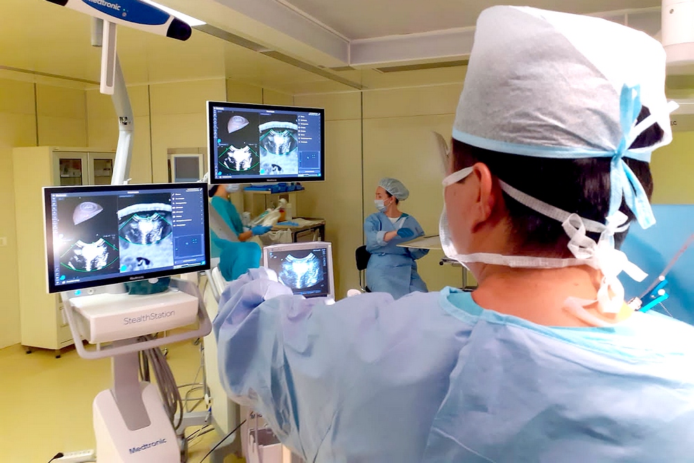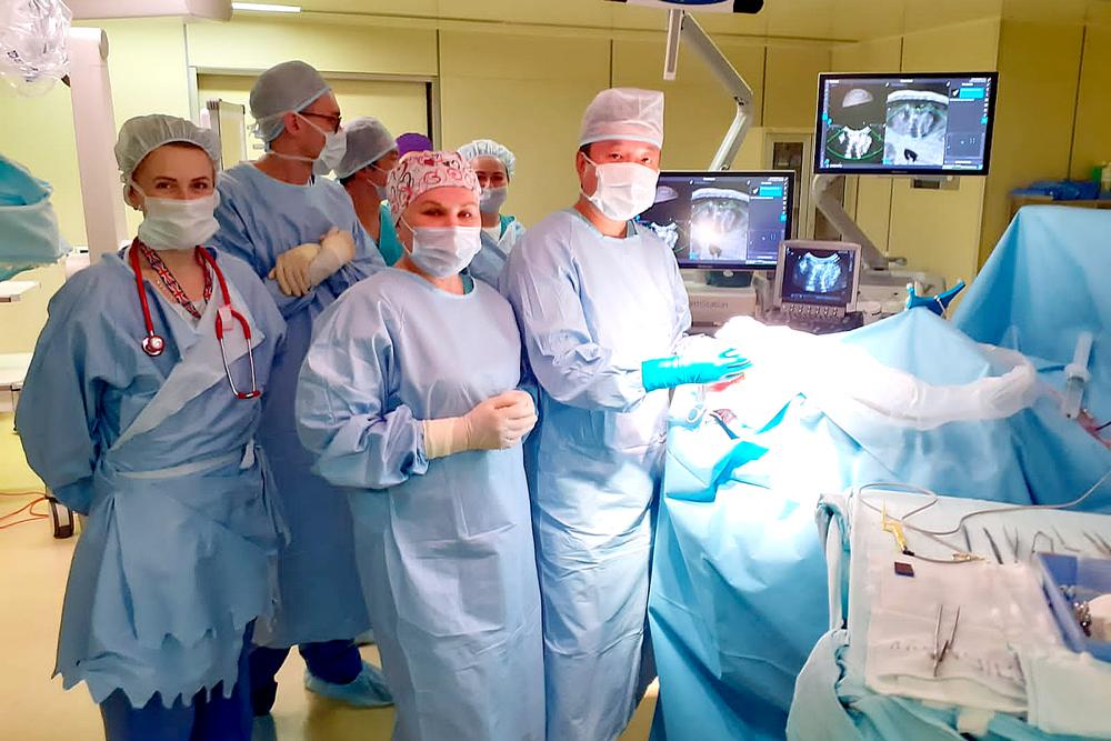
A 15-year-old teenager ended up on the operating table in Pediatric Neurosurgery Department No. 7 at Almazov Centre six months after he was diagnosed with a brain tumor. After a thorough examination of the patient, the doctors decided that the tumor had to be removed by surgery.
Such surgery is usually performed using a neuronavigation system with preloaded patient's MRI data. Thanks to a special program for mapping the optimal path to the tumor, which is sometimes located in the deepest corners of the brain, neurosurgeons were able to get to it with minimal damage to the surrounding tissues.
However, surgery is often associated with a change in the anatomy of the brain, which can cause an error in preoperative coordinates and the chosen trajectory patters.
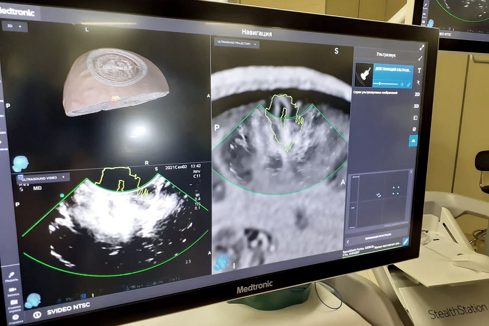
For better accuracy during tumor removal, doctors supplemented intraoperative diagnostic imaging with neurosonography.

“Here at Almazov Centre, we were the first in Russia to simultaneously use the digital integration of an ultrasound and neuronavigation system. This technology makes it possible to combine images of the sonogram and preoperative MRI during surgery (in real time), which significantly improves the interpretation of the neuroimaging picture, making it as complete as possible for full navigation at all stages. Both methods complement each other,” says Alexander Kim, Chief of Pediatric Neurosurgery Department No. 7.
Combining these two technologies into a single diagnostic unit made it possible to minimize the weaknesses while taking maximum advantage of their strengths. By using this technology, doctors managed to completely remove the brain tumor and obtain an encouraging treatment result.
The boy is now doing well and is preparing to be discharged. The tumor was found to be benign. Considering that it was completely removed, in the future he will only have to undergo a control MRI.

