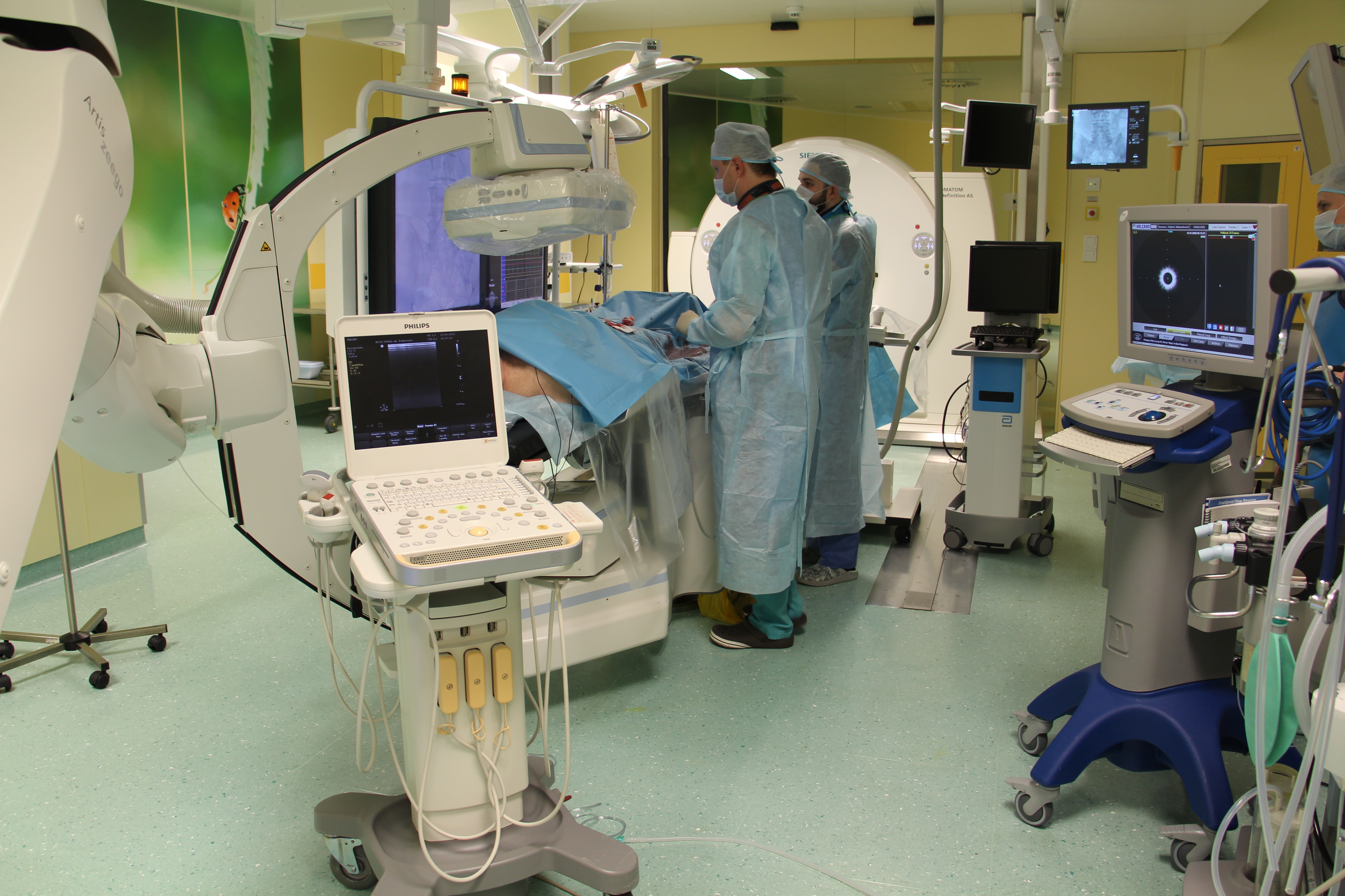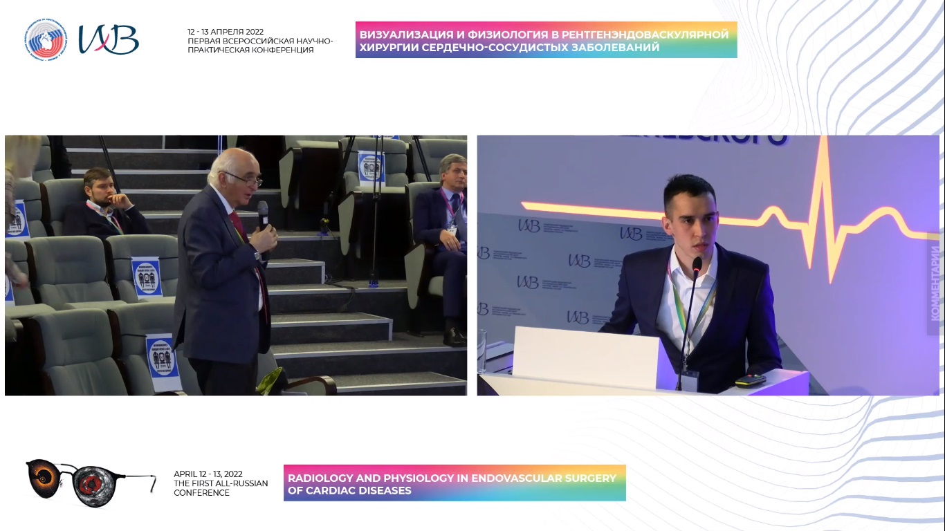
On April 12-13, Vishnevsky National Medical Research Centre of Surgery in Moscow held the first all-Russian conference on Radiology and Physiology in Endovascular Surgery of Cardiac Diseases organized by the Russian Scientific Society of Specialists in X-ray Endovascular Diagnosis and Treatment. More than 300 Russian and foreign endovascular and vascular surgeons, cardiologists, sonographers, specialists in MRI, MSCT and nuclear medicine took part in the event. The conference featured 10 presentations at the plenary session, 50 presentations at breakout sessions and the expert meeting on endovascular methods of diagnosis and treatment.
The conference started with a welcome address by Academician of the Russian Academy of Sciences Bagrat Alekyan and Director of Vishnevsky National Medical Research Centre of Surgery Amiran Revishvili. Academician Alekyan noted that the current rapid development of endovascular surgery is inextricably linked to the constant improvement of imaging methods in the treatment of cardiovascular diseases. In today’s world, a vascular surgeon must master all surgical techniques and be able to interpret the results of all types of vascular imaging in order to achieve the most effective treatment outcomes.

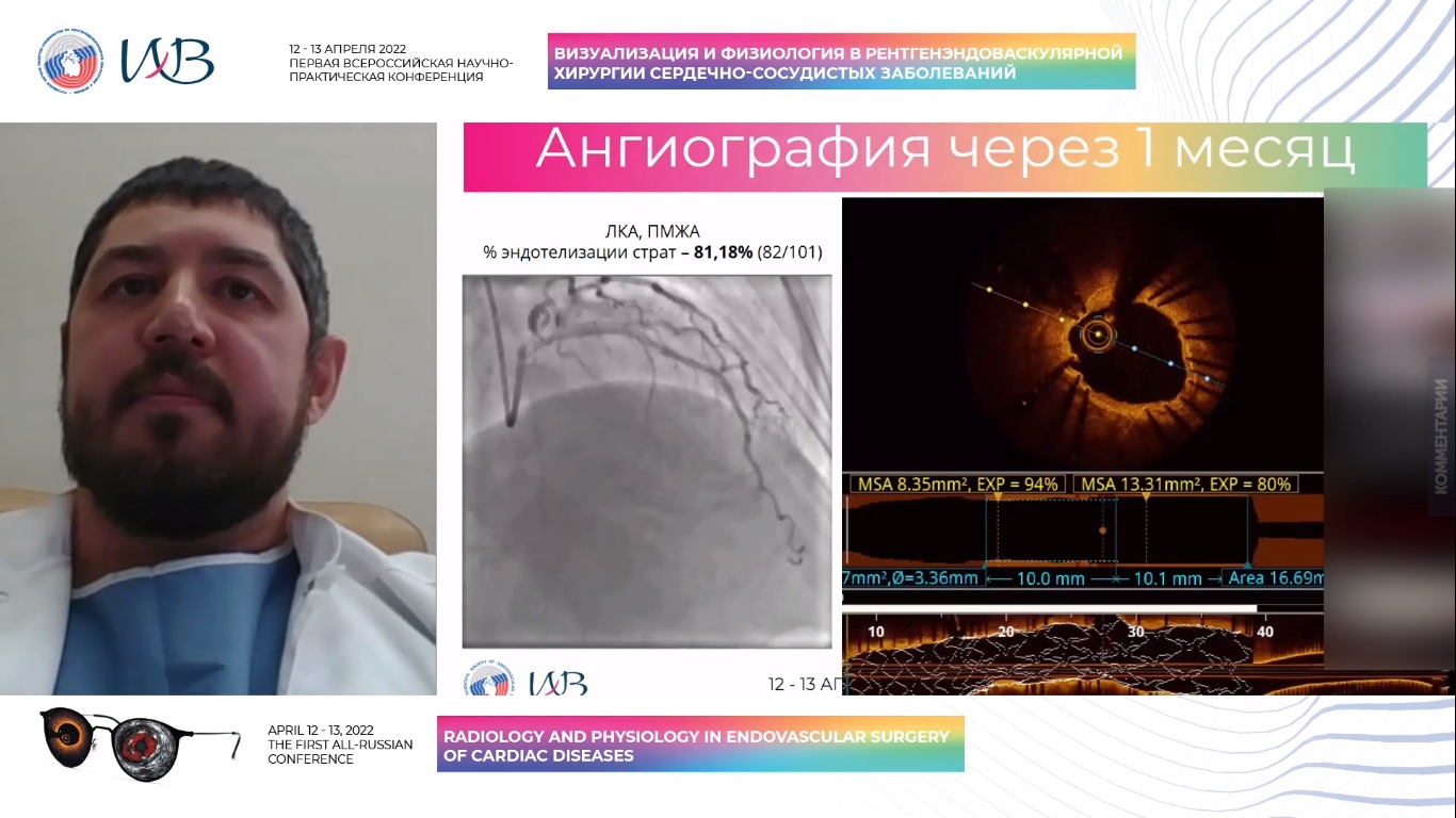
The specialists of the Research Department of Vascular and Interventional Surgery were among the first in Russia to actively introduce methods of intravascular imaging in the treatment of coronary and peripheral arteries. Dmitry Zubarev, Head of the Department of X-ray Surgical Methods of Diagnosis and Treatment, and Almaz Vanyurkin, Researcher at the Research Laboratory of Vascular and Hybrid Surgery, spoke about the unique experience of the clinic in the use of optical coherence tomography (OCT) and intravascular ultrasound (IVUS).

Dmitry Zubarev emphasized the decisive role of OCT in assessing the degree of stent endothelialization when deciding on the early cancellation of dual antiplatelet therapy. This clinical situation occurs in patients with a high risk of bleeding, and therefore additional imaging techniques help choose the optimal treatment tactics for patients with coronary artery disease.
Researcher Almaz Vanyurkin compared various types of imaging in the brachiocephalic artery disease. Academician Bagrat Alekyan highlighted the importance and uniqueness of this study and its practical relevance for Russian specialists in non-standard clinical cases.
Intravascular imaging for peripheral artery interventions is a poorly studied topic but it has critical implications for the treatment of atherosclerosis. In addition to traditional angiography, IVUS and OCT provide additional hemodynamic parameters and data on the nature of atherosclerotic plaque. Undoubtedly, these methods will improve the assessment of the degree and extent of damage to the arterial bed, increase the accuracy of the selection of an implantable device and improve the immediate and long-term outcomes.
The Research Department of Vascular and Interventional Surgery of Almazov Centre headed by Mikhail Chernyavsky continues to actively introduce high-tech modalities into daily clinical use.
