Radiology Unit
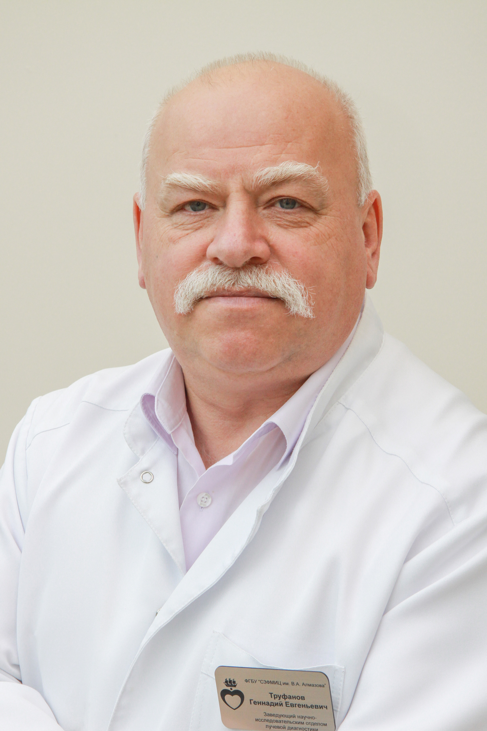 Prof. Gennady Trufanov, MD, DSc
Chief Researcher
History
The Radiology Unit was established in 2009 by optimizing and transforming the Research Laboratory of X-Ray Computed Tomography.
The Radiology Unit comprises:
- Radiological Imaging Laboratory
- Research Group for Radiological Methods in Perinatology and Pediatrics
- MRI Laboratory
Major objectives
- Improvement of diagnostic radiology services for the examination of cardiology, neurology and neurosurgery patients as well as in emergency conditions
- Introduction of new promising diagnostic modalities of magnetic resonance imaging, computed tomography and positron emission tomography.
Major areas
- Development of innovative radiological imaging methods in the field of cardiology (cardiovascular imaging)
- Development and introduction into clinical practice of radiological methods in neurology and neurosurgery (neuroimaging)
- Development of innovative technologies for the study of neural networks in the brain and artificial intelligence
- Introduction into practice of new promising diagnostic methods of magnetic resonance imaging in obstetrics and gynecology
- Development and clinical use of X-ray diagnostic units in neonatology and pediatrics
- Training of research staff.
Training and education
The specialists of the department carry out teaching activities at the Department of Radiology and Medical Imaging of the Medical Education Institute in all areas of study: specialist training, PhD training and residency, continuing professional education.
- PhD – Diagnostic Radiology / Radiation Therapy.
- Residency – X-Ray, Ultrasound.
- Continuing professional education – various professional retraining and advanced training courses.
1. A number of techniques and devices have been developed, improved and implemented for clinical use – the MR hysterosalpingography algorithm for the diagnosis of infertility in women of reproductive age by performing a one-time comprehensive MR study.
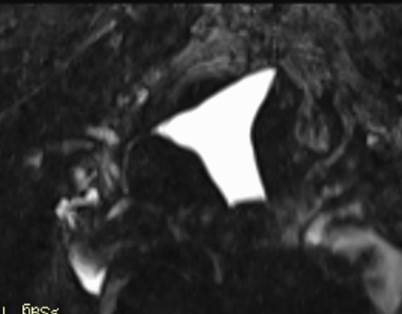
The left fallopian tube is obstructed in the intramural area (arrow), the right fallopian tube is patent (dotted arrow).
2. Together with the LETI scientists, a modern diagnostic X-ray unit for newborns has been improved.
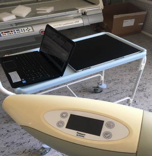
PARDUS X-Ray Diagnostic System for newborns and infants.
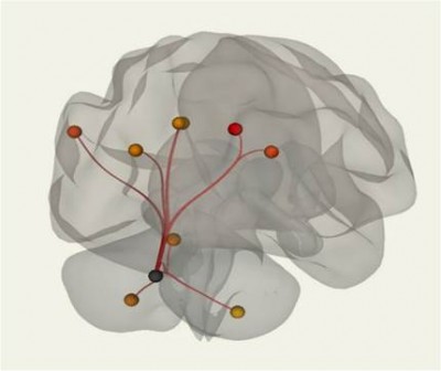
3. Research of the structural and functional state of the brain in various non-tumor and tumor diseases.
 |
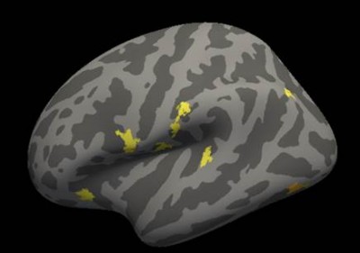 |
| Connectome of the brain. Resting-state cerebellar network | Statistical processing of data on cerebral cortex thickness in patients with multiple sclerosis. |
4. The researchers from the Radiology Unit, together with the scientists from ITMO University were the first in the world to prove the effectiveness of wireless signal transmission in clinical tasks of magnetic resonance imaging. At the same time, they were able to obtain MR images that are not inferior and even superior in quality to those obtained on scanners using signal transmission via radio frequency cables. The findings have been published in Magnetic Resonance in Medicine, the most respected international journal in MRI.
5. Joint research has been carried out in the field of artificial intelligence: the development of a deep neural network for the complete automatic segmentation of the cartilage tissue in the wrist joint, allowing for measuring its thickness, volume, location and severity of changes, which are extremely difficult for visual assessment, even by an experienced radiologist.
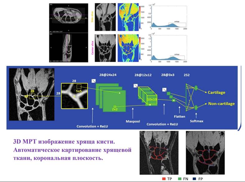
Cartilage segmentation algorithm
State Assignments
Currently, the department is carrying out priority research under four state assignments:
- Development of a new technology for functional neuroimaging to identify early morpho-functional markers in postresuscitation encephalopathy and determine the ways of patient rehabilitation (supervised by A. Efimtsev).
- Development and implementation of a new method of magnetic resonance hysterosalpingography for infertility (supervised by G. Trufanov).
- Innovative digital X-ray diagnostic system for neonatology and pediatrics (supervised by G. Trufanov).
- Development and implementation of a new method for non-invasive determination of iron concentration in the liver and myocardium by magnetic resonance T2 mapping to assess the effectiveness of iron chelation therapy and plan bone marrow transplantation (supervised by G. Trufanov).
Grants
- Methods for analyzing unstructured big data to develop a system for assessing the prognosis for the recovery of integrative brain function and create treatment methods in conditions of impaired consciousness – a combination of loss and new pathological integration of the body (to be completed in May 2022).
- Structural and functional brain changes in patients with chronic insomnia and their relationship with molecular markers of the nervous system function and cardiovascular risk factors (completed in 2019).
Efimtsev A.Y., Levchuk A.G., Trufanov G.E., Kondratyeva E.N., Shmedyk N.Y., Ignatova T.S., Sarana A.M., Shcherbak S.G., Trufanov A.G., Danilov Y.P. Translingual neurostimulation in late residual stage cerebral palsy children treatment affects functional brain networks. HEALTHINF 2019 — 12th International Conference on Health Informatics, Proceedings; Part of 12th International Joint Conference on Biomedical Engineering Systems and Technologies, BIOSTEC2019. 2019;549-556.
Shelepin, KY; Trufanov, GE; Fokin, VA; Vasil'ev, PP; Sokolov, AV. Digital visualization of the activity of neural networks of the human brain before, during, and after insight when images are being recognized. JOURNAL OF OPTICAL TECHNOLOGY. 2018; 85 (8): 468-475. DOI: 10.1364/JOT.85.000468.
Martynov BV, Kholyavin AI, Nizkovolos VB, Parfenov VE, Trufanov GE, Svistov DV. Stereotactic Cryodestruction of Gliomas. Prog Neurol Surg. 2018;32:27-38. DOI: 10.1159/000469677.
Shchelokova, AV; van den Berg, CAT; Dobrykh, DA; Glybovski, SB; Zubkov, MA; Brui, EA; Dmitriev, DS; Kozachenko, AV; Efimtcev, AY; Sokolov, AV; Fokin, VA; Melchakova, IV; Belov, PA. Volumetric wireless coil based on periodically coupled split-loop resonators for clinical wrist imaging. MAGNETIC RESONANCE IN MEDICINE. 2018; 80 (4): 1726—1737. DOI: 10.1002/mrm.27140.
Patent No. 2716750, “Method for treating patients with panic disorders”
Authors: M.L. Pospelova, T.M. Alekseeva, N.E. Ivanova, G.E. Trufanov, V.A. Fokin, A.Yu. Efimtsev, O.A. Chernyshova, A.G. Levchuk, D.N. Iskhakov, N.N. Semibratov
Patent No. 2585403, “Method for assessing the success of osteoporosis treatment”
Authors: D.Yu. Anokhin, G.E. Trufanov, E.N. Tsygan, V.S. Dekan, V.N. Malakhovsky, R.M. Akiev, T.V. Podlesnaya
